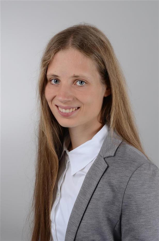

My Research Group uses translational MR imaging to understand how cortical microstructure links to human brain function in health and disease. We study healthy younger and older adults, people with neurodegenerative diseases and people with mental disorders to understand the neuronal mechanisms that underlie healthy and pathological brain states and their modification.
Neuronal Mechanisms & MR Methods Development
My group uses and develops novel MR imaging methods to precisely describe functional and structural circuits in the living human brain. We combine 3T, 7T and 9.4T MRI with novel methods of computational modeling to understand and investigate neuronal mechanisms that underlie human brain pathology and human brain plasticity. We use these methods in our basic science projects but also in clinical projects on aging, neurodegeneration and mental health (see below).
More information on associated projects and publications:
Aging & Neurodegeneration
We use a combination of 3T, 7T and 9.4T MRI, behavioral assessments, neurophysiological measurements and automatic tools to better understand human brain aging in health and disease. Our focus is to investigate degenerating sensory and motor brain circuits that play a major role for everyday wellbeing, and are impaired in various neurodegenerative disorders such as motor neuron disease or Parkinson Disease. New and more sensitive imaging and analyses methods aid early diagnosis and help to better understand the underlying neuronal mechanisms, such as neuroinflammation, substance accumulation, or disease spread. This forms the basis for an individualized neuromedicine, where therapeutic interventions are tailored to the individual patient.
More information on associated projects and publications
Mental Health
A major challenge for modern neuroscience research is to understand the neuronal mechanisms that underlie mental health. Understanding these mechanisms may allow us to maintain mental health in vulnerable populations, and to stabilize it in situations of disbalance. Body memories have a major influence on our daily behavior and play a particularly important role for processing traumatic experiences. We investigate human body memory in a multimodal approach by using 3T, 7T and 9.4T MRI and also integrate novel tools of virtual reality (VR) to experimentally induce and investigate the complex processes that give rise to human body memories. These insights provide evidence-based information that can be used to inform and develop novel psychosomatic and therapeutic interventions.

+49 (0)7071-
92-54151
Complete list of publication on ORCID
Selected publications
Northall A, Doehler J, Weber M, Tellez I, Petri S, Prudlo J, Vielhaber S, Schreiber S, Kuehn E (2024) Multimodal layer modelling reveals in-vivo pathology in ALS. Brain 147:1087-99
Northall A, Doehler J, Weber M, Vielhaber S, Schreiber S, Kuehn E (2023) Layer-Specific Vulnerability is a Mechanism of Topographic Map Aging. Neurobiol Aging 128:17-32
Doehler J, Northall A, Liu P, Fracasso A, Chrysidou A, Speck O, Lohmann G, Wolbers T, Kuehn E (2023) The 3D Structural Architecture of the Human Hand Area is Non-Topographic. J Neurosci 43:3456-3476
Gentsch A, Kuehn E (2022) Clinical Manifestations of Body Memories: The Impact of Past Bodily Experiences on Mental Health. Brain Sciences 12:594
Liu P, Chrysidou A, Doehler J, Wolbers T, Kuehn E (2021) The organizational principles of de-differentiated topographic maps in somatosensory cortex. eLife 10:e60090
Schreiber S, Northall A, Weber M, Vielhaber S, Kuehn E (2021) Topographic layer-imaging as a tool to track neurodegenerative disease spread in M1. Nat Rev Neurosci 22:68-69
Lohmann G, Stelzer J, Lacosse E, Kumar VJ, Müller K, Kuehn E, Grodd W, Scheffler K (2018) LISA improves statistical analyses for fMRI Nat Commun 9:4014
Kuehn E, Sereno I M (2018) Modelling the human cortex in three dimensions Trends Cogn Sci 22:1073-1075.
Kuehn E, Haggard P, Villringer A, Pleger B, Sereno M (2018). Visually-driven maps in area 3b. J Neurosci 38:1295-1310.
Kuehn E, Dinse J, Jacobsen E, Villringer A, Sereno M, Margulis D (2017) Body topography parcellates human sensory and motor cortex. Cereb Cort 10:1-16.
Open positions for the research group can be found here:
https://www.estherkuehn-science.org/open-positions.html
Cooperations with Groups within the HIH
- FG Giese (neuronal mechanisms of real life movements)
- FG Kowarik (neuronal immune profiling of MS patients)
- FG Snaidero (combined human-mice investigation of cortical lesions)
- FG Lerche (layer-specific lesion detection in epilepsy patients)
- FG Synofzik (imaging of human neurodegeneration)

Hertie Institute for Clinical Brain Research
Otfried-Müller-Straße 27
72076 Tübingen


