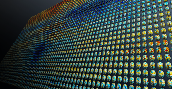

The central goal of our laboratory is to investigate how cognition and behavior emerges from dynamic interactions across widely distributed neuronal ensembles. How do sophisticated cognitive processes such as perception, decision-making, and motor behavior emerge from dynamic interactions across the brain? Which neural mechanisms coordinate these interactions, how are they dynamically regulated in a goal-directed fashion, and how are these interactions disturbed in neuropsychiatric diseases?
To address these questions, we apply an interdisciplinary multi-scale approach and link large-scale population measures of neuronal activity to circuit and cellular-level mechanisms. To this end, we combine human MEG and EEG, animal electrophysiology, psychophysics and sophisticated analytical techniques.
Our research is funded by the German Research Foundation (DFG) and European Research Council (ERC).


Neuronal Information through Neuronal Interactions (NINI)
Our thoughts and actions arise from the activity of specialized neurons in the brain. Much is already known about which neurons and brain regions encode sensory, cognitive or motor information. However, comparatively little is known about which specific interactions of neurons establish this information. This is because neuronal interactions and information coding have been studied in largely separate research areas. The central aim of the NINI project is to bridge this gap.
To this end we combine state-of-the-art electrophysiological and analytical techniques. Magnetoencephalography (MEG) is used to measure the brain activity in humans during decision-making. We investigate which interactions between brain areas underlie the decisions made and the neuronal coding of information. Furthermore, we investigate the link between circuit interactions and information coding during decision-making at the level of individual neurons. From this multi-scale approach and the linking of two largely separate fields, we expect to gain new insights into the neuronal basis of our thinking and acting.
This project is funded by the European Research Council (ERC) under the European Union’s Horizon 2020 research and innovation programme (ERC Consolidator Grant; Grant agreement No. 864491).

Spectral Fingerprints of Normal and Diseased Brain Function
One focus of the lab are oscillatory dynamics of neuronal activity. Brain activity exhibits oscillations, i.e. periodicity, at various different frequencies and spatial scales. These oscillations may not only serve as highly informative markers, or ‘spectral fingerprints’ of the circuit interactions involved in different cognitive functions but may also dynamically mediate these interactions.
In one line of research, we investigate these spectral fingerprints with MEG in the resting human brain. We investigate how networks of brain regions spontaneously coordinate their oscillations at different frequencies. Together with our clinical collaborators, we investigate these rhythmic networks as novel biomarkers of different neurological pathologies. Furthermore, we characterize the temporal microstructure of different brain rhythms and how they are linked to neuronal spiking at the cellular level.
Neuronal Dynamics During Cognition and Behavior
In another line of research, we investigate neural dynamics underlying specific cognitive functions. We characterize the flow of information and neuronal interactions across distributed cortical and sub-cortical networks during complex behavioral tasks, involving e.g. visual and auditory decision-making, attention, working memory, and proprioception. We investigate how sensory, cognitive, and motor neuronal information flows across the brain and how such information relates to neuronal oscillations, dynamics and interactions. Together with our clinical collaborators, we investigate alterations of neuronal dynamics during pathological conditions, such as e.g. dyslexia in children and spatial hearing in cochlear implant users.
Tübingen Magnetoencephalography (MEG) Center – Imaging Human Brain Dynamics
Magnetoencephalography (MEG) allows for non-invasively and directly measuring human brain activity with unparalleled temporal resolution and signal quality. Our lab operates the Tübingen MEG Center, which provides state-of-the-art MEG techniques and services to the Tübingen neuroscience community and beyond. The MEG Center hosts a 275-channel whole-head SQUID-based MEG system, synchronized high-density EEG, transcranial electrical stimulation, precise visual, auditory and somatosensory stimulation, various response systems and high-speed binocular eye-tracking. Furthermore, the MEG-Center employs novel optically pumped magnetometers (OPMs) to measure brain activity.
The MEG Center is involved in a broad spectrum of collaborative research projects with partners at the Center for Neurology, the Centre for Integrative Neuroscience, the Institute of Medical Psychology, the Department of Otolaryngology, the Centre for Ophthalmology, the Department of Psychiatry and Psychotherapy and the Department of Psychosomatic Medicine.
Magnetomyography (MMG) – Quantum Sensing of Muscle Activity
Quantum sensors are well-established to record brain activity, i.e., for Magnetoencephalography (MEG), but not for studies of muscles, i.e., magnetomyography (MMG). In contrast to the electrical pendant, i.e, electromyography (EMG), MMG has the advantage that it can be recorded without contact and that the surrounding tissues only minimally affect the propagation of magnetic signals generated by the muscles. This offers the possibility of contactless, high-fidelity measurements of muscle activity for a broad spectrum of applications. In this line of research, our group investigates the physiological principles of MMG and its application for neurological diagnostics, neurorehabilitation and prosthesis control using different quantum sensors, such as optically pumped magnetometers (OPMs), Nitrogen-vacancy (NV) based magnetometers and superconductive quantum interference devices (SQUID).


Selected publications
· Pellegrini F, Hawellek DJ, Pape AA, Hipp JF, Siegel M (2020) Motion Coherence and Luminance Contrast Interact in Driving Visual Gamma-Band Activity. Cerebral Cortex ePub
· Siems M, Siegel M (2020) Dissociated neuronal phase- and amplitude-coupling patterns in the human brain. Neuroimage 209:116538
· Trainito C, von Nicolai C, Miller EK, Siegel M (2019) Extracellular spike waveform dissociates four functionally distinct cell classes in primate cortex. Current Biology 29
· Fioravanti C, Kajal SD, Carboni M, Mazzetti C, Ziemann U, Braun C (2019) Inhibition in the somatosensory system: An integrative neuropharmacological and neuroimaging approach. Neuroimage; 202:116139.
· Sandhaeger F, von Nicolai C, Miller EK, Siegel M (2019) Monkey EEG links neuronal color and motion information across species and scales. eLife 8:e45645
· Wang H, Braun C, Murphy EF, Enck P (2019) Bifidobacterium longum 1714™ Strain Modulates Brain Activity of Healthy Volunteers During Social Stress. Am J Gastroenterol; 114(7):1152-1162
· Broser PJ, Knappe S, Kajal DS, Noury N, Alem O, Shah V, Braun C (2018) Optically Pumped Magnetometers for Magneto-Myography to Study the Innervation of the Hand. IEEE Trans Neural Syst Rehabil Eng; 26(11): 2226-2230.
· Pape AA and Siegel M (2016) Motor cortex activity predicts response alternation during sensorimotor decisions. Nature Communications 7:13098
· Siegel M, Buschman TJ and Miller EK (2015) Cortical Information Flow During Flexible Sensorimotor Decisions. Science 348(6241): 1352-55
· Hipp JF and Siegel M (2015) BOLD fMRI Correlation Reflects Frequency-Specific Neuronal Correlation. Current Biology 25, 1368-1374
· Weisz N, Wühle A, Monittola G, Demarchi G, Frey J, Popov T, Braun C. (2014). Prestimulus oscillatory power and connectivity patterns predispose conscious somatosensory perception. PNAS.;111(4): E417-425.
· Hipp JF, Hawellek DJ, Corbetta M, Siegel M and Engel AK (2012) Large-scale cortical correlation structure of spontaneous oscillatory activity. Nature Neuroscience 15:884-890
· Siegel M, Donner TH and Engel AK (2012) Spectral fingerprints of large-scale neuronal interactions. Nature Reviews Neuroscience 13:121-134
· Hipp JF, Engel AK and Siegel M (2011) Oscillatory Synchronization in Large-Scale Cortical Networks Predicts Perception. Neuron 69: 387-396
· Siegel M, Warden MR and Miller EK (2009) Phase-Dependent Neuronal Coding of Objects in Short-Term Memory. PNAS 106(50): 21341–21346
· Siegel M, Donner TH, Oostenveld R, Fries P and Engel AK (2008) Neuronal Synchronization along the Dorsal Visual Pathway Reflects the Focus of Spatial Attention. Neuron 60: 709-719
· Miltner WHR, Braun C, Arnold M, Witte H, Taub E (1999) Coherence of gamma-band EEG activity as a basis for associative learning. Nature, 397: 434-436.

Hertie Center of Neurology
Hertie Institute for Clinical Brain Research
Department of Neural Dynamics and Magnetoencephalography
Otfried-Müller-Straße 25
72076 Tübingen
Phone: +49 (0)7071 29-85297
Fax: +49 (0)7071 29-25002


